

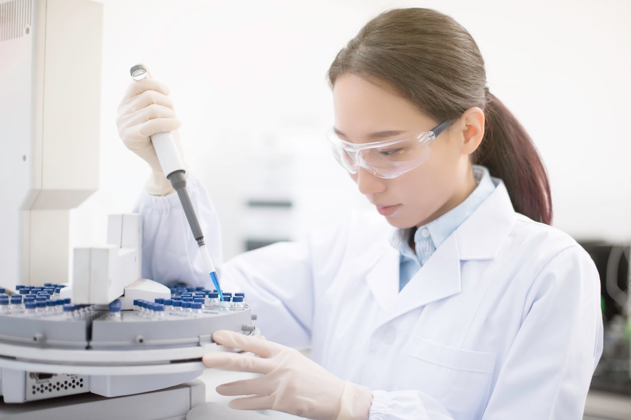
A biopsy is a medical procedure used to remove a small sample of cells, tissue, or sometimes fluid from the body so it can be examined in a laboratory. It is one of the most important diagnostic tools in modern medicine, particularly when doctors need to determine whether an abnormal area is benign, precancerous, or cancerous. The type of biopsy recommended depends on several factors, including the location and size of the abnormality, the suspected condition, and the patient's overall health.
Why Is a Biopsy Performed?
Doctors may recommend a biopsy when imaging tests, physical examinations, blood tests, or other diagnostic procedures reveal an abnormality that requires closer examination. A biopsy can provide information that imaging alone cannot. Biopsies are commonly used to investigate:
Suspicious lumps or masses
Abnormal skin lesions
Breast abnormalities
Lung nodules
Liver abnormalities
Enlarged lymph nodes
Abnormalities in the prostate
Gastrointestinal lesions
Abnormal bone or soft tissue
Unexplained changes in certain organs
A biopsy does not necessarily mean that cancer is suspected. Although biopsies are widely associated with cancer diagnosis, they can also be used to identify infections, inflammatory diseases, and other non-cancerous conditions.
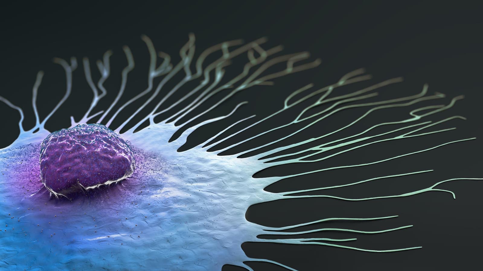
What Are the Different Types of Biopsy?
There are several types of biopsy. They can generally be categorized according to how the tissue sample is collected and whether a needle, endoscope, surgical procedure, or other technique is used.
Needle Biopsy
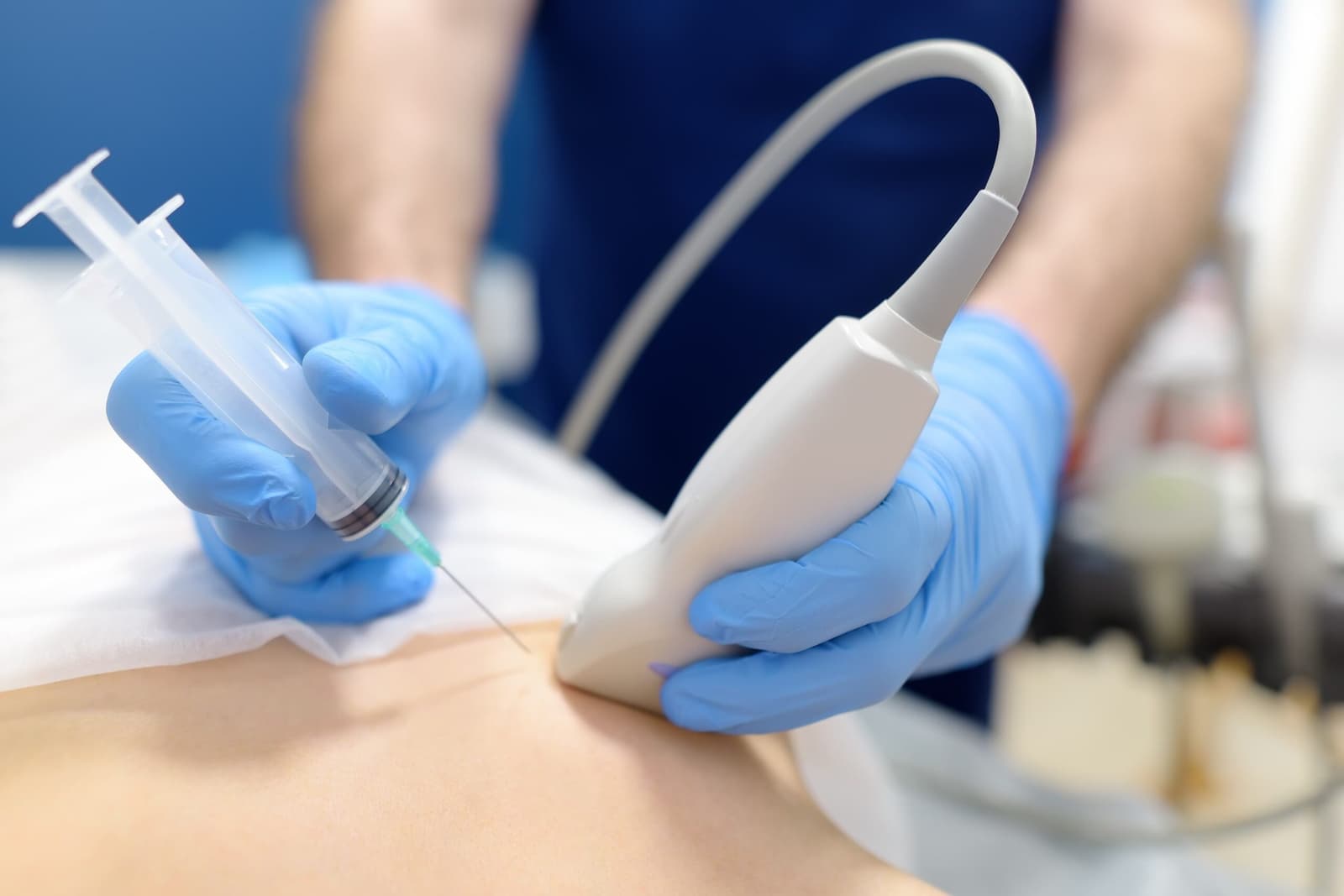
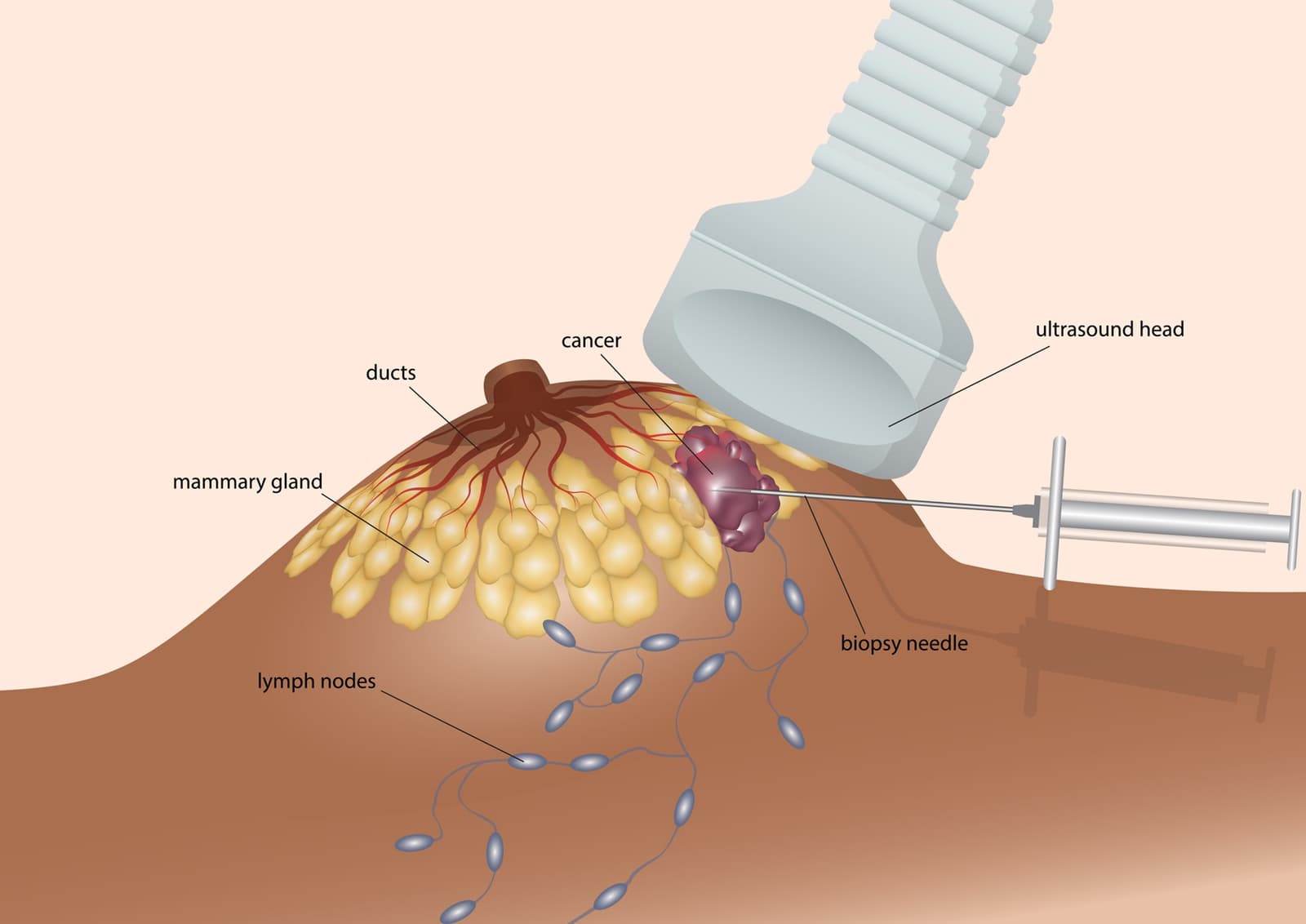
Needle biopsy is one of the most common biopsy techniques. It involves inserting a needle through the skin and into the area being investigated to collect cells or tissue. Needle biopsies are often performed with the assistance of imaging technologies such as ultrasound, CT, or mammography to help the doctor accurately target the abnormal area.
There are several forms of needle biopsy, including fine-needle aspiration and core needle biopsy.
Fine-Needle Aspiration (FNA)
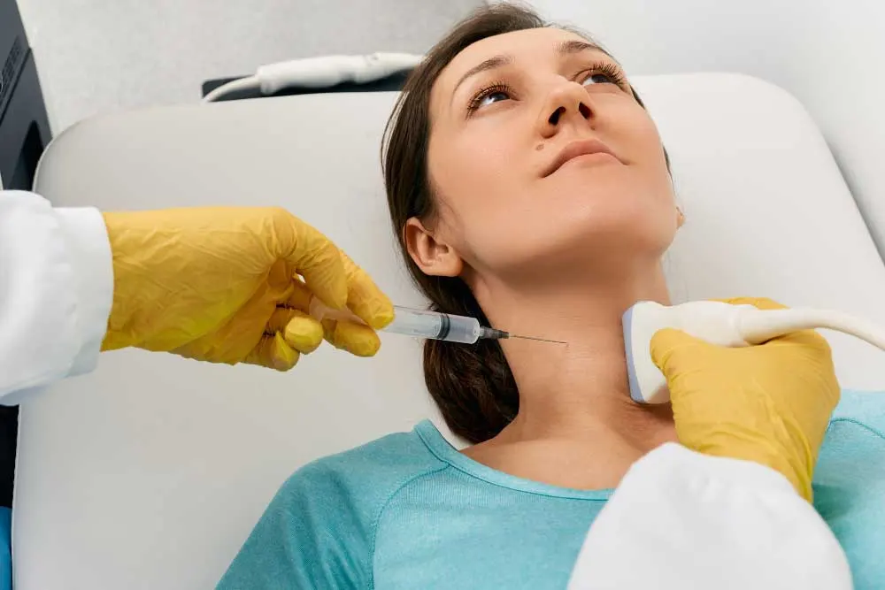
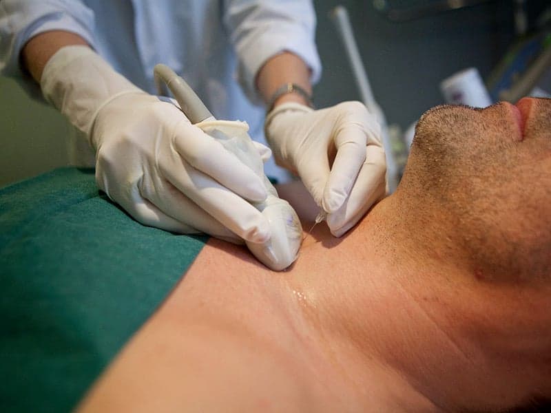
Fine-needle aspiration uses a very thin needle to remove a small number of cells or fluid from a suspicious area. The sample is then examined under a microscope. FNA is frequently used to evaluate lumps in areas such as the thyroid, lymph nodes, salivary glands, and breasts. It is generally a minimally invasive procedure and may only require local anesthesia or, in some cases, no anesthesia.
One limitation is that FNA collects individual cells rather than a larger piece of tissue. Therefore, depending on the suspected condition, the doctor may recommend a core needle biopsy instead.
Core Needle Biopsy
A core needle biopsy uses a larger hollow needle to remove small cylinders, or "cores," of tissue. Because it collects a larger and more structured tissue sample than FNA, it can provide additional information about the architecture of the tissue. Core needle biopsies are commonly used for breast, liver, prostate, and other soft-tissue abnormalities. Imaging guidance may be used to ensure the needle reaches the correct location.
Core biopsy is often performed under local anesthesia and usually does not require a hospital stay.
Vacuum-Assisted Biopsy
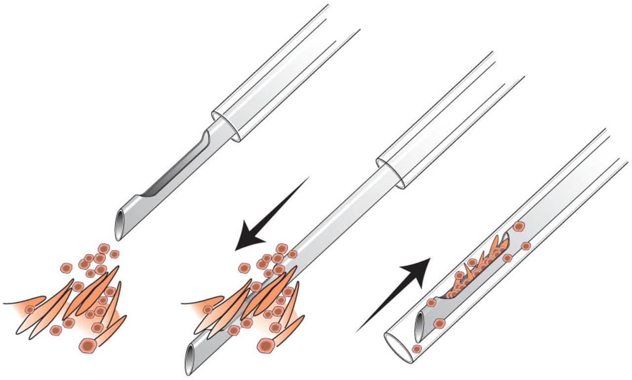
A vacuum-assisted biopsy uses a specialized needle connected to a vacuum device. The vacuum helps draw tissue into the needle, allowing multiple samples to be collected through a single skin opening. This technique is particularly useful for certain breast abnormalities and can sometimes remove a larger portion of a small lesion than a conventional needle biopsy.
Vacuum-assisted biopsy is generally minimally invasive and is often performed with imaging guidance.
Incisional Biopsy
An incisional biopsy is a surgical procedure in which only a portion of an abnormal area or lesion is removed for examination. This method may be appropriate when an abnormality is too large to remove completely or when the doctor needs to establish a diagnosis before planning definitive treatment.
An incisional biopsy can be performed under local or general anesthesia depending on the location and size of the lesion.
Excisional Biopsy
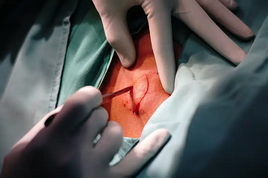
An excisional biopsy involves removing the entire suspicious lesion or abnormal area, often along with a small margin of surrounding tissue. This technique may be used for certain skin lesions, small lumps, or other accessible abnormalities. In some situations, removing the entire lesion provides both a diagnosis and treatment.
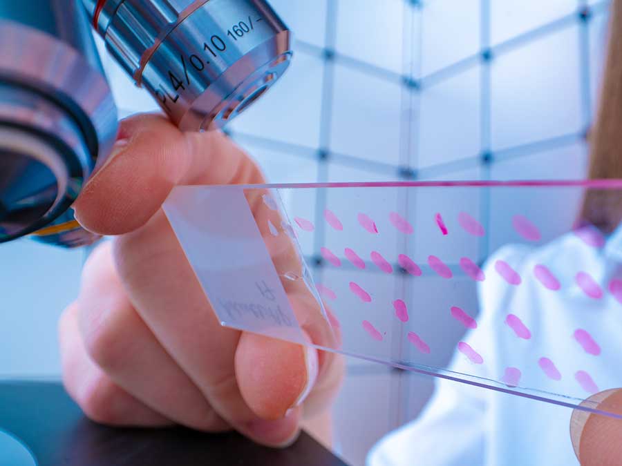
The removed tissue is sent to a pathology laboratory, where a specialist examines it for abnormal or cancerous cells.
Punch Biopsy
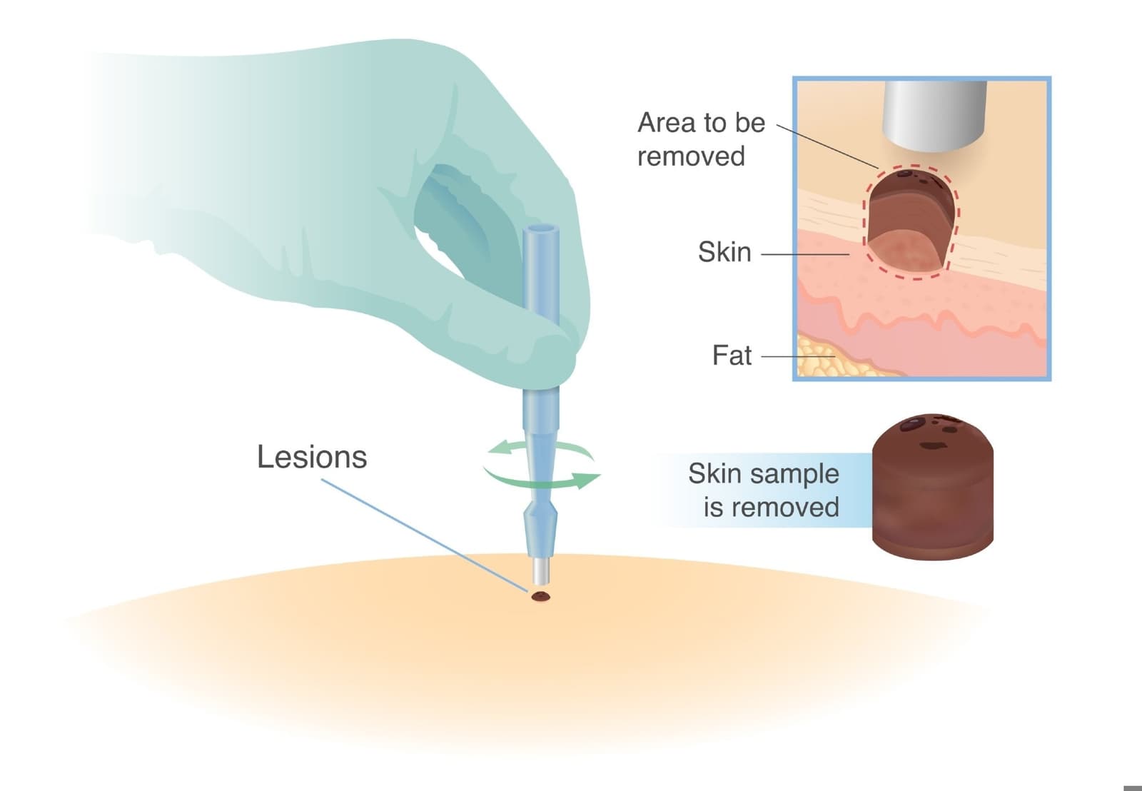
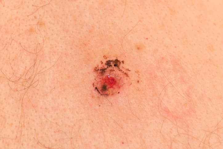
A punch biopsy is commonly used to examine abnormalities of the skin. During the procedure, the doctor uses a small, circular cutting instrument to remove a cylindrical section of skin containing several layers of tissue. Punch biopsies can help diagnose conditions such as suspicious skin lesions, inflammatory skin diseases, and certain types of skin cancer.
The procedure is usually performed with local anesthesia. Depending on the size of the sample, the wound may require one or more stitches.
Shave Biopsy
A shave biopsy involves removing the upper layers of an abnormal skin lesion using a blade or similar instrument. It is generally used for superficial skin abnormalities and is less invasive than procedures that remove deeper layers of tissue. A shave biopsy may be suitable for certain raised or superficial skin lesions.
However, because it does not collect deeper tissue, it may not be appropriate for every suspicious lesion. The doctor chooses the biopsy technique based on the characteristics and suspected depth of the abnormality.
Endoscopic Biopsy
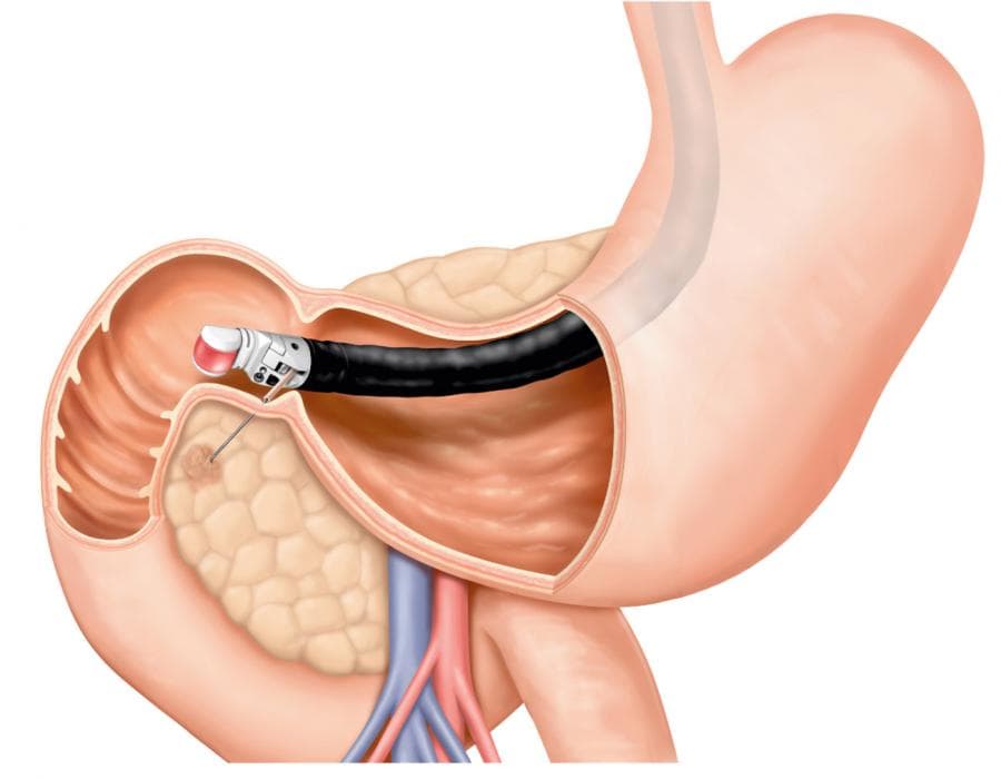
An endoscopic biopsy is performed during an endoscopy. An endoscope is a flexible or rigid instrument equipped with a camera that allows doctors to examine the inside of certain organs. During the procedure, specialized instruments can be passed through the endoscope to collect tissue samples.
Endoscopic biopsies may be performed during procedures such as upper gastrointestinal endoscopy or colonoscopy. They can help investigate abnormalities in the esophagus, stomach, small intestine, colon, and other areas accessible through an endoscope.
Bone Biopsy
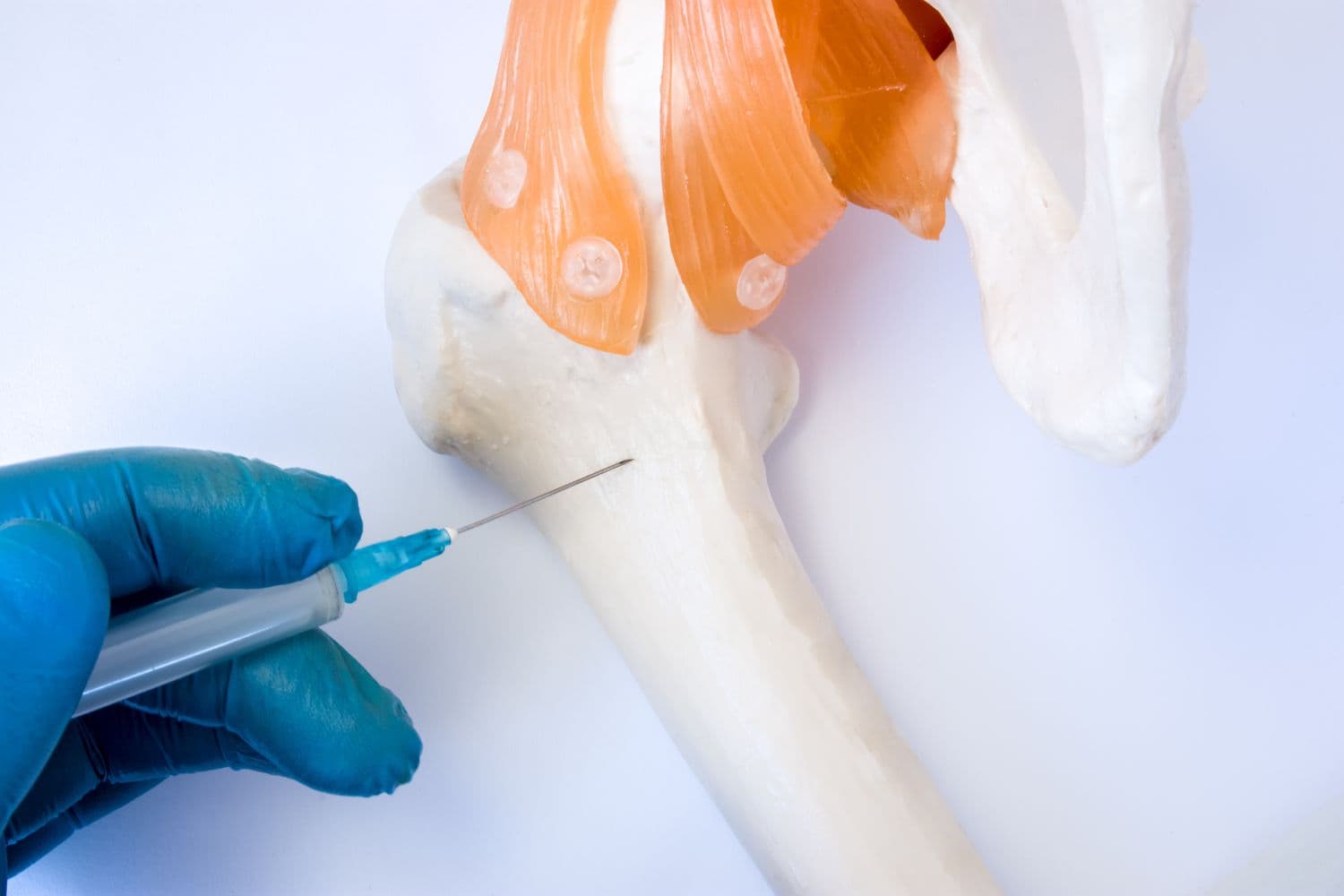
A bone biopsy involves collecting a sample of bone tissue for laboratory examination. It may be recommended when doctors need to investigate an unusual bone lesion, unexplained bone changes, or a suspected bone tumor.
Bone biopsies may be performed using a needle or through a surgical procedure, depending on the location and characteristics of the abnormality. Imaging guidance is often used for needle-based procedures.
Bone Marrow Biopsy
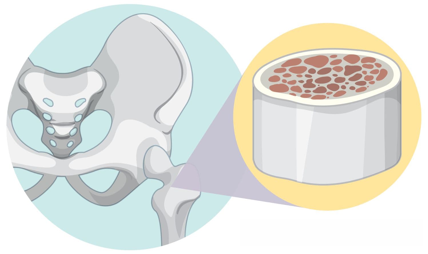
A bone marrow biopsy is different from a bone biopsy. It examines the soft tissue inside certain bones where blood cells are produced. The procedure commonly involves collecting bone marrow from the back of the pelvic bone. Doctors may recommend a bone marrow biopsy to investigate blood disorders, unexplained abnormalities in blood counts, leukemia, lymphoma, or other conditions affecting blood-cell production.
A bone marrow examination may involve both aspiration, which collects liquid marrow, and a core biopsy, which collects a small solid piece of marrow-containing bone.
Lymph Node Biopsy
A lymph node biopsy involves removing cells or tissue from a lymph node for laboratory examination. It may be recommended when a lymph node is enlarged or has an unusual appearance. Depending on the circumstances, doctors may use FNA, core needle biopsy, or surgical removal of part or all of a lymph node.
Lymph node biopsies can be important in diagnosing infections, inflammatory diseases, and cancers that may involve the lymphatic system.
How Is the Right Biopsy Type Selected?
There is no single biopsy technique that is appropriate for every patient. Doctors consider the location, size, depth, and appearance of the abnormality, as well as the suspected disease. Imaging tests can help determine which technique is safest and most effective. For example, ultrasound or CT guidance may be used to accurately target an internal lesion, while a skin abnormality may be examined using a punch, shave, incisional, or excisional biopsy.
The expected information from the biopsy is also important. Some conditions require an intact piece of tissue so that the pathologist can examine the structure of the cells and surrounding tissue. In other situations, collecting a small number of cells through FNA may be sufficient.
What Happens After a Biopsy?
After tissue is collected, it is normally sent to a pathology laboratory. A pathologist examines the sample using microscopic and, when necessary, specialized laboratory techniques. The pathology report may determine whether the tissue is normal, benign, inflammatory, infectious, precancerous, or malignant. Additional tests may sometimes be performed on the tissue to identify specific characteristics of abnormal cells.
The time required to receive results varies depending on the type of biopsy and the laboratory tests needed. Patients should discuss the expected timeframe and follow-up process with their healthcare provider.
What Are the Risks of a Biopsy?
Most biopsies are considered safe, but every medical procedure carries potential risks. The risks depend on the type and location of the biopsy. Possible complications include:
Mild pain or discomfort
Bruising or swelling
Bleeding
Infection
Temporary numbness
Scarring
Damage to nearby structures in uncommon cases
More invasive biopsies may have additional risks related to anesthesia or surgery. Before undergoing a biopsy, patients should tell their healthcare team about medications, allergies, bleeding disorders, and other relevant medical information.
Conclusion
Biopsy procedures are essential diagnostic tools that allow doctors to examine abnormal cells and tissues directly. From minimally invasive fine-needle aspiration and core needle biopsy to surgical, endoscopic, skin, bone, and bone marrow biopsies, different techniques are designed for different medical situations.
Read More