

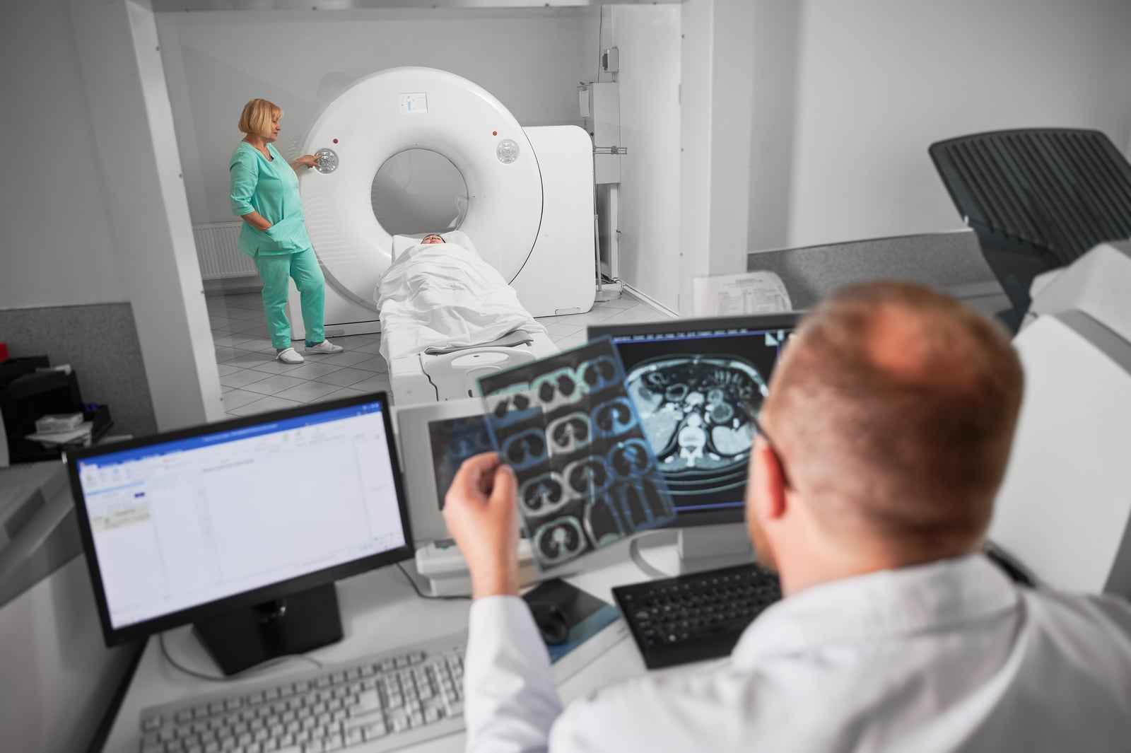
Diagnostic imaging plays a vital role in modern medicine, allowing healthcare providers to look inside the human body without the need for invasive procedures. From detecting fractures to diagnosing complex conditions like cancer or neurological disorders, imaging technologies provide precise and detailed insights. Each imaging method works differently and involves unique steps, equipment, and preparation requirements.
X-ray: What Happens During the Procedure?
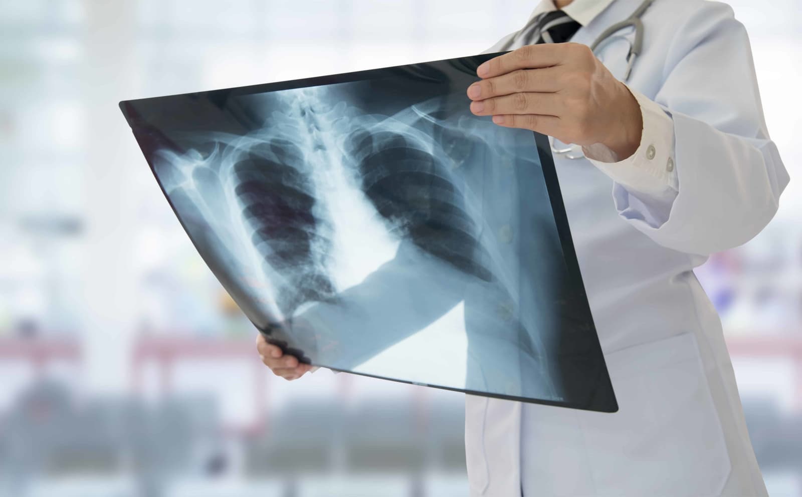
An X-ray is one of the most common and widely used imaging techniques. It uses a small amount of radiation to create images of the inside of the body, particularly bones and dense structures. During an X-ray procedure, the patient is typically asked to stand, sit, or lie down depending on the area being examined. A radiologic technologist positions the body carefully to capture the correct angle. In some cases, multiple images may be taken from different positions to ensure accuracy. Patients are often asked to remain completely still for a few seconds while the image is captured. Movement can blur the image, making it harder to interpret. The process itself is quick and painless, usually taking only a few minutes.
In certain cases, patients may be asked to wear a lead apron to protect other parts of the body from radiation exposure. The amount of radiation used is minimal and generally considered safe. Once the images are taken, they are reviewed by a radiologist who looks for abnormalities such as fractures, infections, or structural issues. Results are often available quickly, making X-rays a fast and efficient diagnostic tool.
CT Scan (Computed Tomography): What Happens During the Procedure?
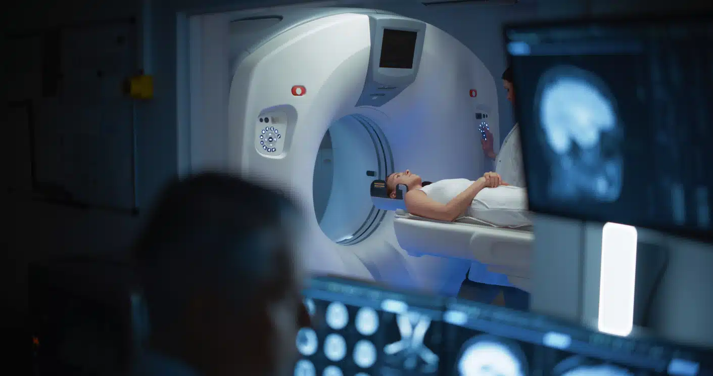
A CT scan is a more advanced imaging technique that uses a series of X-ray images taken from different angles to create detailed cross-sectional images of the body. During a CT scan, the patient lies on a motorized table that slides into a large, donut-shaped machine. The scanner rotates around the body, capturing multiple images in a short period of time. The procedure is painless and typically lasts between 10 to 30 minutes.
In some cases, a contrast dye may be used to enhance the visibility of certain tissues or blood vessels. This dye can be administered orally, intravenously, or occasionally rectally, depending on the area being examined. Patients receiving contrast may feel a warm sensation or a metallic taste, which usually passes quickly. It is important for patients to remain still during the scan to ensure clear images. In certain situations, they may be asked to hold their breath briefly. After the scan, patients can usually resume normal activities immediately unless they were given sedation or contrast dye that requires observation. CT scans are highly effective in diagnosing conditions such as internal injuries, tumors, and infections.
MRI (Magnetic Resonance Imaging): What Happens During the Procedure?
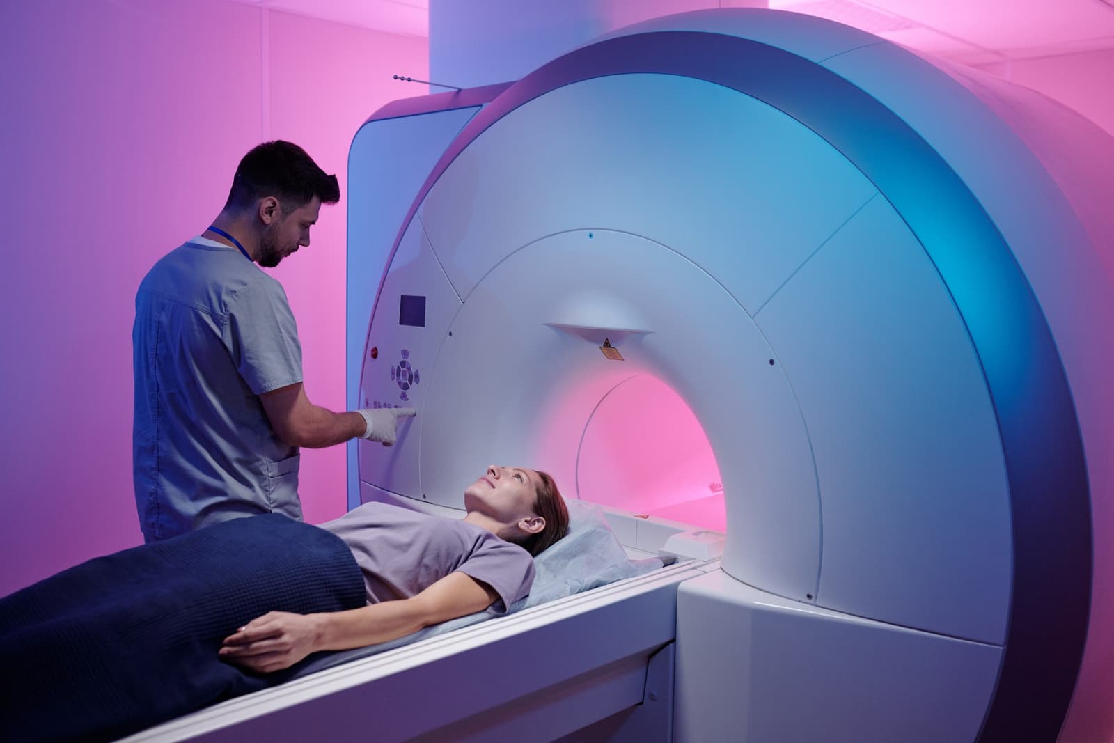
MRI is a powerful imaging technique that uses strong magnetic fields and radio waves to create detailed images of organs and tissues. Unlike X-rays and CT scans, MRI does not use ionizing radiation. During an MRI scan, the patient lies on a table that slides into a long, tube-shaped machine. The procedure can take anywhere from 20 to 60 minutes, depending on the area being examined. Patients are required to remain very still during the scan, as movement can affect image quality. The machine produces loud tapping or knocking noises during operation, so patients are often given earplugs or headphones for comfort.
In some cases, a contrast agent may be injected into a vein to improve image clarity. Patients should inform their doctor of any allergies or kidney issues beforehand. Because MRI uses strong magnets, patients must remove all metal objects, including jewelry, watches, and sometimes even clothing with metal components. It is also important to inform the medical team about any implants, such as pacemakers. MRI is particularly useful for imaging soft tissues, including the brain, spinal cord, muscles, and joints, making it essential for diagnosing neurological and musculoskeletal conditions.
PET Scan (Positron Emission Tomography): What Happens During the Procedure?
A PET scan is a specialized imaging test that helps evaluate metabolic activity in the body. It is often used in oncology, cardiology, and neurology to detect cancer, assess heart function, or study brain disorders. Before the scan, a small amount of radioactive tracer is injected into the patient’s bloodstream. This tracer highlights areas of high metabolic activity, which may indicate disease. After the injection, patients typically wait 30 to 60 minutes to allow the tracer to distribute throughout the body. During this time, they are usually asked to rest quietly.
The scan itself involves lying on a table that moves through a PET scanner, similar in appearance to a CT machine. The procedure is painless and generally takes about 20 to 40 minutes. Patients must remain still during the scan to ensure accurate results. Afterward, they are advised to drink plenty of fluids to help flush the tracer from the body. PET scans are particularly valuable for detecting cancer at an early stage, evaluating treatment effectiveness, and identifying abnormal cellular activity.
Ultrasound: What Happens During the Procedure?
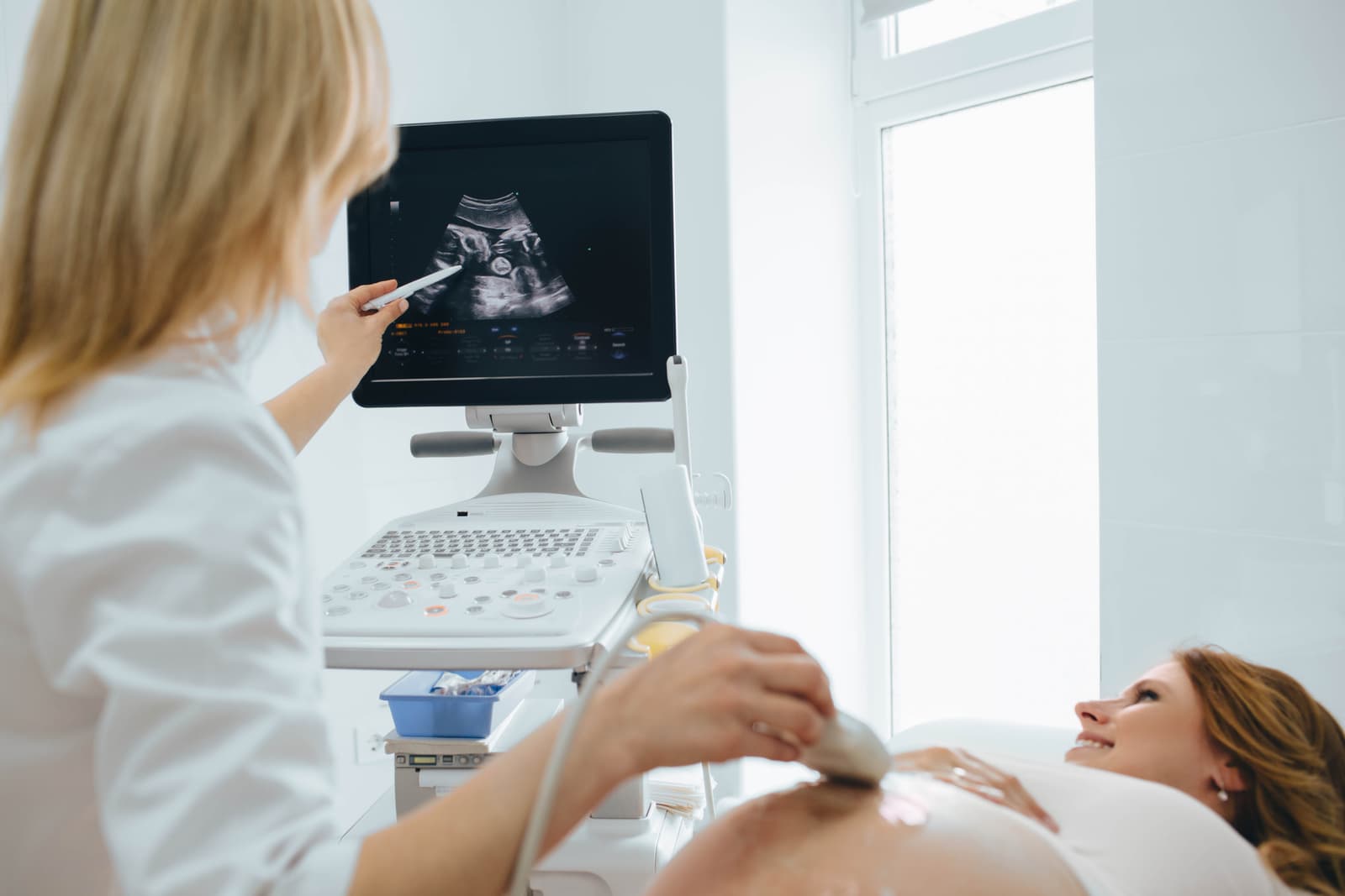
Ultrasound imaging uses high-frequency sound waves to create real-time images of the body’s internal structures. It is a safe and non-invasive method that does not involve radiation. During an ultrasound, the patient typically lies on an examination table while a technician applies a special gel to the skin over the area being examined. This gel helps transmit sound waves and improves image quality. A handheld device called a transducer is then moved over the skin. The transducer emits sound waves that bounce off internal organs and tissues, creating images on a monitor.
The procedure is painless and usually takes between 15 to 45 minutes. Patients may be asked to change positions or hold their breath briefly to improve image clarity. Ultrasound is commonly used to examine the abdomen, pelvis, heart, and developing fetus during pregnancy. It is also useful for guiding certain medical procedures, such as biopsies. Because it is safe, quick, and widely available, ultrasound is often the first imaging test recommended for many conditions.
Conclusion
Diagnostic imaging is an essential part of modern healthcare, providing detailed insights that guide accurate diagnosis and effective treatment. Each imaging method—whether X-ray, CT scan, MRI, PET scan, or ultrasound—offers unique advantages and serves specific medical purposes. Being informed and prepared not only enhances patient confidence but also ensures a smoother and more efficient medical travel experience.
Read More