


Dacryocystorhinostomy, commonly abbreviated as DCR, is a surgical procedure used to treat blocked tear drainage. When tears cannot drain normally from the eye into the nose, they can accumulate in the lacrimal system and cause excessive tearing, recurrent eye infections, discharge, or swelling near the inner corner of the eye.
DCR creates a new pathway between the lacrimal drainage system and the nasal cavity, allowing tears to bypass the blocked portion of the nasolacrimal duct and drain into the nose more effectively. There are two main approaches to DCR: external DCR and endonasal DCR.
What Is Dacryocystorhinostomy?
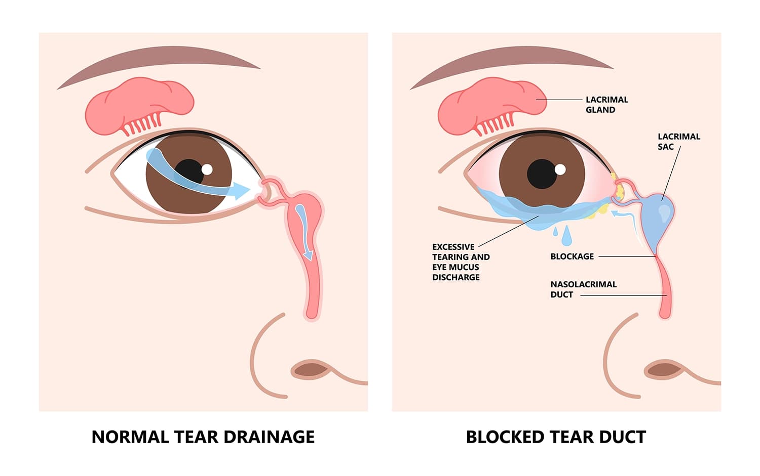
Normally, tears produced by the lacrimal glands drain across the surface of the eye and enter small openings called puncta. From there, tears travel through the canaliculi into the lacrimal sac and then through the nasolacrimal duct into the nasal cavity. If the nasolacrimal drainage pathway becomes narrowed or blocked, tears may accumulate rather than draining normally. This condition is called nasolacrimal duct obstruction. Common symptoms include:
Persistent watery eyes
Tears running down the face
Recurrent eye infections
Discharge or mucus around the eye
Swelling or tenderness near the inner corner of the eye
Blurred vision caused by excessive tearing
When conservative treatments are not sufficient, DCR may be recommended.

What Is External DCR?
External DCR is the traditional approach to creating a new tear drainage pathway. During the procedure, the surgeon makes a small incision in the skin near the inner corner of the eye. The surgeon then accesses the lacrimal sac and creates an opening between the lacrimal sac and the nasal cavity.
The new opening allows tears to drain directly into the nose instead of passing through the obstructed portion of the original drainage pathway. The skin incision is usually positioned within or near a natural skin crease. Although a scar can develop, it is generally designed to be as inconspicuous as possible.
External DCR has been performed for many years and remains an important treatment option for nasolacrimal duct obstruction.
What Is Endonasal DCR?
Endonasal DCR, sometimes called endoscopic DCR, creates the new drainage pathway from inside the nose. Instead of making an external skin incision, the surgeon uses instruments inserted through the nasal cavity. An endoscope provides visualization of the nasal structures and the area surrounding the lacrimal sac. The surgeon creates an opening between the lacrimal sac and the nasal cavity so that tears can drain through the newly created pathway.
Because the operation is performed through the nose, there is no external facial incision. Depending on the patient's anatomy and the surgeon's approach, additional nasal procedures may sometimes be performed at the same time.
External vs. Endonasal DCR: What Is the Difference?
The main difference between external and endonasal DCR is where the surgeon accesses the lacrimal drainage system.
In external DCR, the surgeon reaches the lacrimal sac through a small incision in the skin near the inner corner of the eye. This approach provides direct access to the lacrimal sac and surrounding structures. Because it involves a skin incision, a small external scar is possible. In endonasal DCR, the surgeon reaches the lacrimal system through the nasal cavity using an endoscope. Since there is no external skin incision, this approach does not leave a facial surgical scar. It also allows the surgeon to directly examine the nasal cavity and identify anatomical factors that may contribute to tear drainage problems.
External DCR has a long history of use and provides direct visualization of the lacrimal sac. It may be particularly useful in certain complex cases or when direct access to the lacrimal system is desirable. Endonasal DCR avoids an external incision and may be particularly suitable for patients whose nasal anatomy or conditions can be addressed during the procedure. However, it requires specialized endoscopic equipment and appropriate surgical expertise.
Both techniques are designed to create a new pathway between the lacrimal sac and the nasal cavity, allowing tears to bypass the blocked portion of the normal drainage system. The choice between them depends on factors such as the location and cause of the obstruction, the patient's lacrimal and nasal anatomy, previous surgery, and the surgeon's experience.
Advantages of External DCR
One potential advantage of external DCR is the direct access it provides to the lacrimal sac. This can allow the surgeon to clearly identify the relevant anatomy and create the new drainage pathway.
External DCR has also been extensively studied and has a long history of use for treating nasolacrimal duct obstruction.
Another consideration is that external DCR may be useful in certain complex cases where direct access to the lacrimal sac is desirable.
The main disadvantage is the external incision. Although the resulting scar is often small and placed strategically, some patients may prefer to avoid any visible facial incision.
Advantages of Endonasal DCR
The most obvious advantage of endonasal DCR is the absence of an external skin incision. This means there is no facial surgical scar from the DCR itself.
Endonasal surgery can also allow the surgeon to directly evaluate the nasal cavity and identify anatomical factors that may contribute to the obstruction.
For patients who have relevant nasal conditions, an endonasal approach may provide an opportunity to address certain nasal problems during the same surgical session when clinically appropriate.
However, endonasal DCR requires specialized equipment and expertise in nasal endoscopic surgery. The suitability of this approach depends partly on the patient's anatomy and the surgeon's experience.
Is One Technique More Successful Than the Other?
Both external and endonasal DCR can be effective treatments for nasolacrimal duct obstruction. Research comparing the two approaches has generally found that success rates can be similar when the procedures are appropriately selected and performed by experienced surgeons.
However, success depends on several factors beyond the surgical approach. These may include the location and severity of the obstruction, previous lacrimal surgery, nasal anatomy, scarring, inflammation, and postoperative care.

Recovery After DCR
Recovery varies according to the surgical technique and the individual patient. After DCR, patients may experience temporary swelling, bruising, nasal congestion, mild discomfort, or a small amount of bleeding or discharge. The healthcare team will provide instructions about medication, nasal care, and activities during recovery.
Some patients may have a temporary silicone tube or stent placed within the tear drainage system. The purpose is to help maintain the new drainage pathway while tissues heal. The timing of stent removal varies according to the patient's condition and the surgeon's treatment plan.
Possible Risks and Complications
DCR is generally considered a well-established procedure, but complications can occur. Potential risks include:
Bleeding
Infection
Swelling or bruising
Scarring
Persistent tearing
Recurrent blockage
Failure of the new drainage pathway to remain open
Nasal discomfort or congestion
Problems related to a temporary stent
External DCR also carries the possibility of a visible skin scar, although the incision is generally designed to minimize its appearance.
Conclusion
Dacryocystorhinostomy is an established surgical treatment for blocked tear drainage. External DCR reaches the lacrimal system through a small incision near the inner corner of the eye, while endonasal DCR approaches it through the nasal cavity without an external skin incision. Both approaches can provide effective relief from symptoms caused by nasolacrimal duct obstruction.
Read More